Ocular Echography
What is Ocular Echography?
It’s a current and useful diagnostic method for eye-related health problems. Traditional echographic studies are essential, for instance, for the the discovering of ocular structures in case of diptric media opacity, the observation and evaluation of the iridocorneal angle amplitude and the evaluation of the ocular musculature. Thanks to its most innovative applications, it is possible to use the echographic sign for the measurement of the eyeball, an essential step in the measurament of the crystalline lens for cataract surgery.

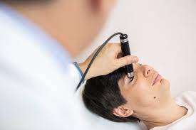
This test does not present any risk and it can be repeated more times; it consists in leaning on the eyelids a tool that does an ultrasound scan of the deep tissues.
It does NOT need any surgical cut or injection.
It does NOT need anesthesia.
- Compared to other techniques, it is not traumatic nor dangerous for the health, since it does not use ionizing radiation, nor contrast medium.
- It is a tomographic technique since images rebuild sections of the tissue.
- It is a “dynamic ” method, since during execution it is possible to evaluate not only on fixed images but also on the moving ones, asking the patient to move the look and moving the echographic probe.
B-SCAN ECHOGRAPHY
Ecography B-SCAN with B-SCAN PLUS®
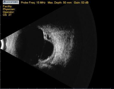
B-SCAN ECHOGRAPHY is a non-invasive diagnostic examination that gives detailed bi-dimensional images of the eyeball and the orbit in order to localize and configure lesions.
It is the most important diagnostic test in the presence of opacity in the dioptric media (advanced cataract) to get information about intraocular structures, differently from the other techniques (to detect either retinal detachment or vitreal hemorrhages).
It is useful also in the presence of transparent dioptric media in the differential diagnosis and the evaluation of intraocular tumors dimension.
Regarding orbital level, it allows to evaluate the expansive lesions (tumors), the extraocular muscle (as in the case of thyroid diseases) and the optic nerve in ist infraorbital nerve.

Our device
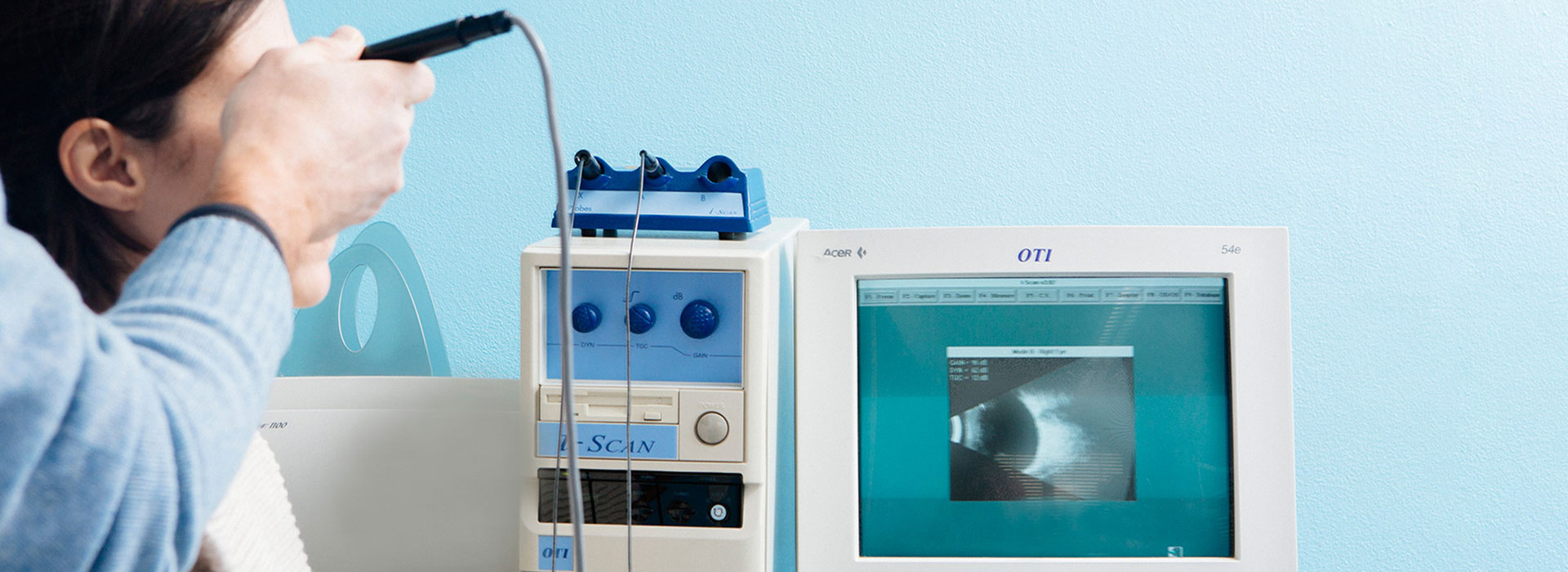
Our latest B-SCAN PLUS® portable probe is a practical tool that can be plugged into any laptop or pc.
In addition to the high resolution image (15 micron), with the loss of signal removal as in the common ecographies, and to the possibility to take lasting 34 seconds shot, in order to determine in vivo the histologic aspect of lesions, our device is extremely maneuverable and we can print or send via e-mail the medical report. Its Smooth Zoom technology allows for 2x full image zoom without distortion or loss of resolution, making this device unique and avantgarde.
Benefits
It’s a painless non-invasive detection that can be performed comfortably with an outpatient service.
It is vital for the children study, even the youngest, since it can be performed without them being sedated and exposed to radiation. For this reason, ecography is ideal for the follow-up of the lesions that need several check-up.
